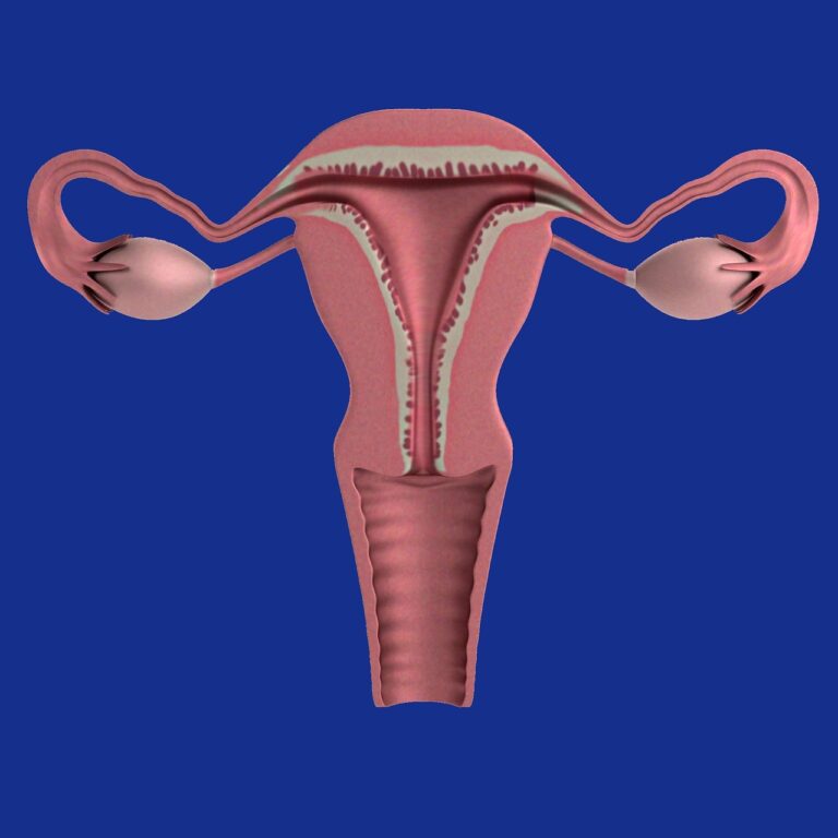
Adenomyosis, a benign uterine condition, is related to endometriosis, and represents the presence of endometrial glands and stroma within the uterine myometrium. The uterus is an organ made up of two types of tissues: endometrium and myometrium.
The pathogenesis of adenomyosis remains elusive, and no single existing theory can explain all disease phenotypes.
Adenomyosis is considered a specific entity in the PALM-COEIN FIGO classification of causes of abnormal uterine bleeding as follows (3):
PALM – polyp; adenomyosis; leiomyoma; malignancy and hyperplasia;
COEIN – coagulopathy; ovulatory dysfunction; endometrial; iatrogenic and not yet classified;
FIGO – International Federation of Gynecology and Obstetrics;

Causes
As with endometriosis, there are several theories explaining how the muscle becomes invaded by the endometrial lining.
So, according to one of these theories, adenomyosis may be caused by direct invasion of the endometrium into the immediately adjacent myometrium. Another possibility is that the muscle tissue for some reason turns into another tissue that resembles the endometrial lining. This process is called metaplasia, the change of one type of tissue to another.
Another theory, which is also the most well-known, says that adenomyosis occurs as a result of the extension of deep pelvic endometriosis from the outside (abdominal-pelvic cavity, peritoneum) to the thickness of the uterine wall (myometrium). This almost always happens in cases of advanced deep endometriosis, when the endometriosis “nodule” infiltrates the posterior uterine wall (cervix, uterine body).
Adenomyosis is classified as diffuse and focal.
- Diffuse adenomyosis represents invasion of endometrial glands and/or stroma within the myometrium;
- Focal adenomyosis or adenomyoma, circumscribed tumors consisting of endometrium and muscle tissue;
Symptoms
- Heavy or prolonged menstruation;
- Extremely painful menstruation;
- Pain during intercourse;
- Clots in menstrual blood;
- Uterus voluminous and sensitive;
- General pain in the pelvic area;
- Sensation of pressure on the bladder and rectum;
- Painful stool;
- Pain during urination;
- Abnormal vaginal bleeding;
- Feeling sick;
- Infertility;
- Abortive disease
Diagnostic
Suspicion of adenomyosis can be made with the help of imaging methods, but also following the vaginal cough that can reveal an enlarged uterus, and sensitive to palpation. However, the final diagnosis of adenomyosis is made after the histological examination of surgically removed specimens. Imaging can consist of transvaginal ultrasound and MRI.
Treatment methods
Treatment of adenomyosis is divided into medical therapy and surgical therapy, which can be conservative or radical, depending on the type of disease and whether fertility is to be preserved.
The surgical treatment of choice for focal adenomyosis is to remove the nodules by excision. Focal excision of adenomyosis can be performed if the location of the lesions can be determined. The procedure can be performed by laparoscopy, mini-laparotomy or laparotomy.
The radical surgical therapy of choice in the case of diffuse adenomyosis consists of hysterectomy which can be total or subtotal, a procedure that is not indicated in the case of patients who wish to become pregnant.
